Investigation of these patients includes thorough clinical examination, audiological evaluation and frequently Magnetic Resonance Imaging (MRI) of the Internal Auditory Meatus (IAM), cerebellopontine angle (CPA) and brain. MRI scanning is a well-established, cost-effective investigation for these patients [1-3]. Only a small percentage of these.. MRI is firmly established as an essential modality in the imaging of the temporal bone and lateral skull base. It is used to evaluate normal anatomic structures, evaluate for vestibular schwannomas, assess for inflammatory and/or infectious processes, and detect residual and/or recurrent cholesteatoma. It is also extensively used in pre- and postoperative evaluations, particularly in patients.
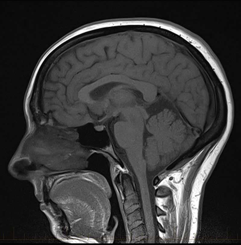
Brain Scans Mri

Normal BRAIN MRI image YouTube

MRI Reveals Striking Brain Differences in People With Autism Brain scan, Mri brain

MRI Scans What to Expect at Moffitt Cancer Center YouTube

BrainMriWithContrastShowingmsimages Mri scan, Mri, Scan

Normal brain MRI Radiology Case Brain images, Mri, Brain scan
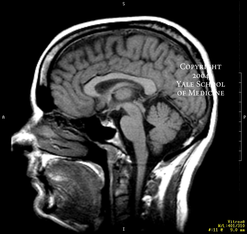
22C+ October 2010
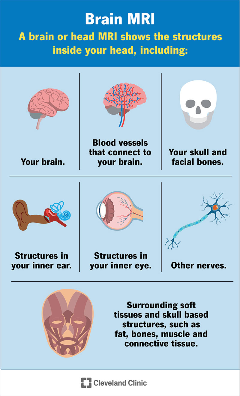
Top 18 what is an mri scan used to diagnose 2022
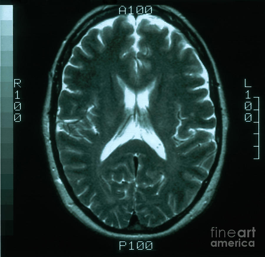
Female Normal MRI Brain
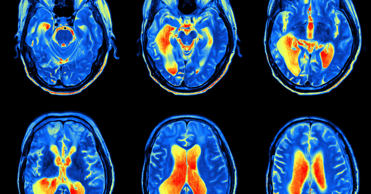
MRI scan image of brain Regional Medical Imaging

Signs of Intracranial Hypertension on MRI Not Commonly Associated With Papilledema Neurology

The Highest Resolution MRI Scan of a Human Brain
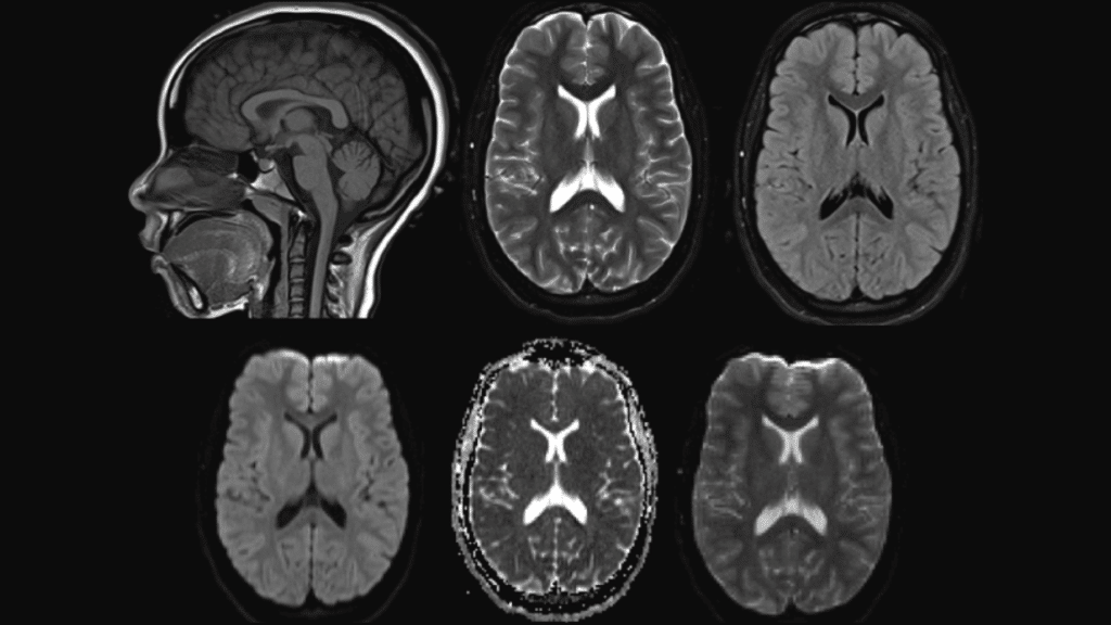
CT vs. MRI What's the Difference? Windom Area Health

Picture Of Brain Mri Machine machinejuli
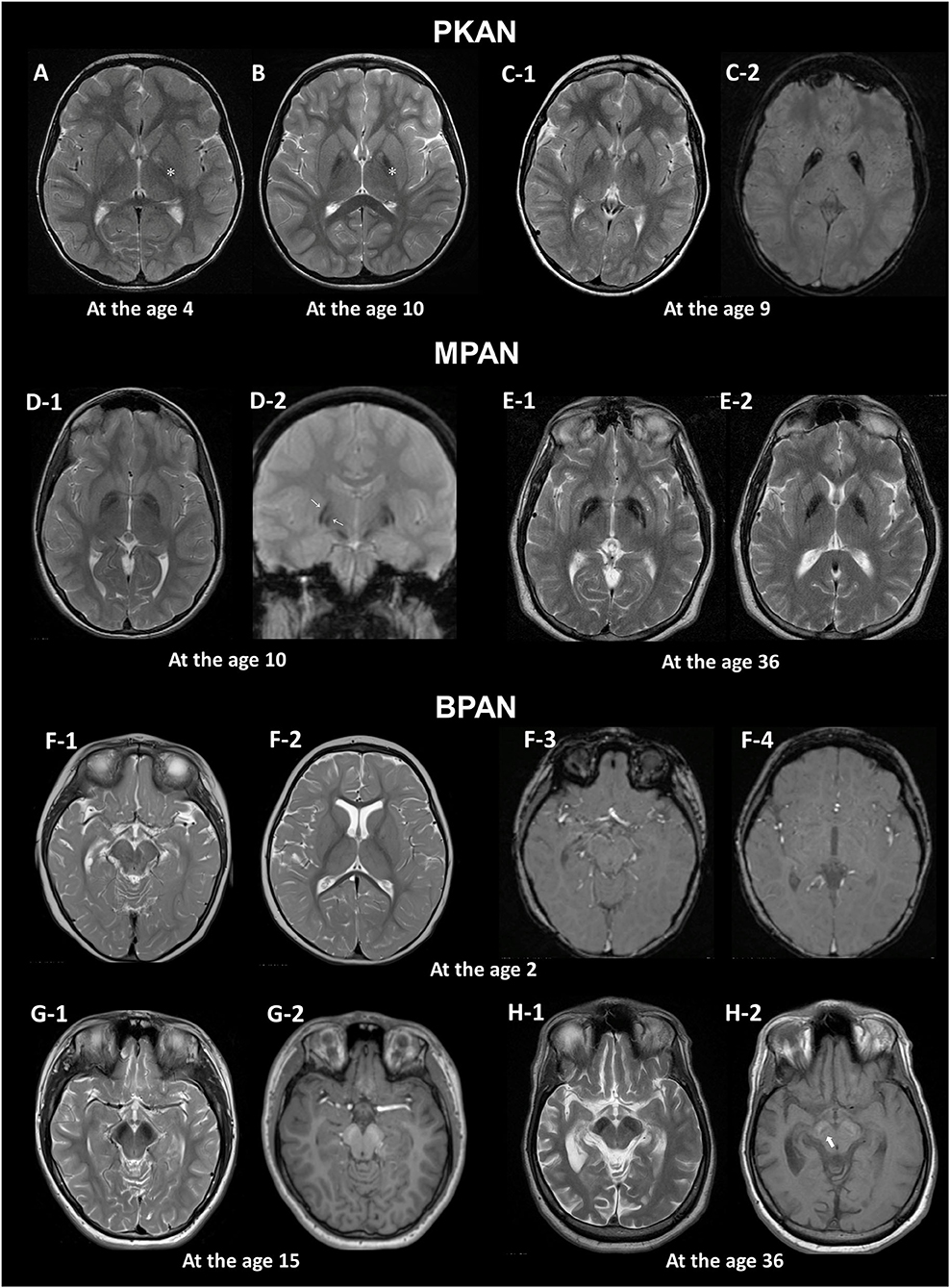
Mri Abnormal Brain Scan

Learn mri brain

Brain MRI scan protocols, positioning and planning YouTube
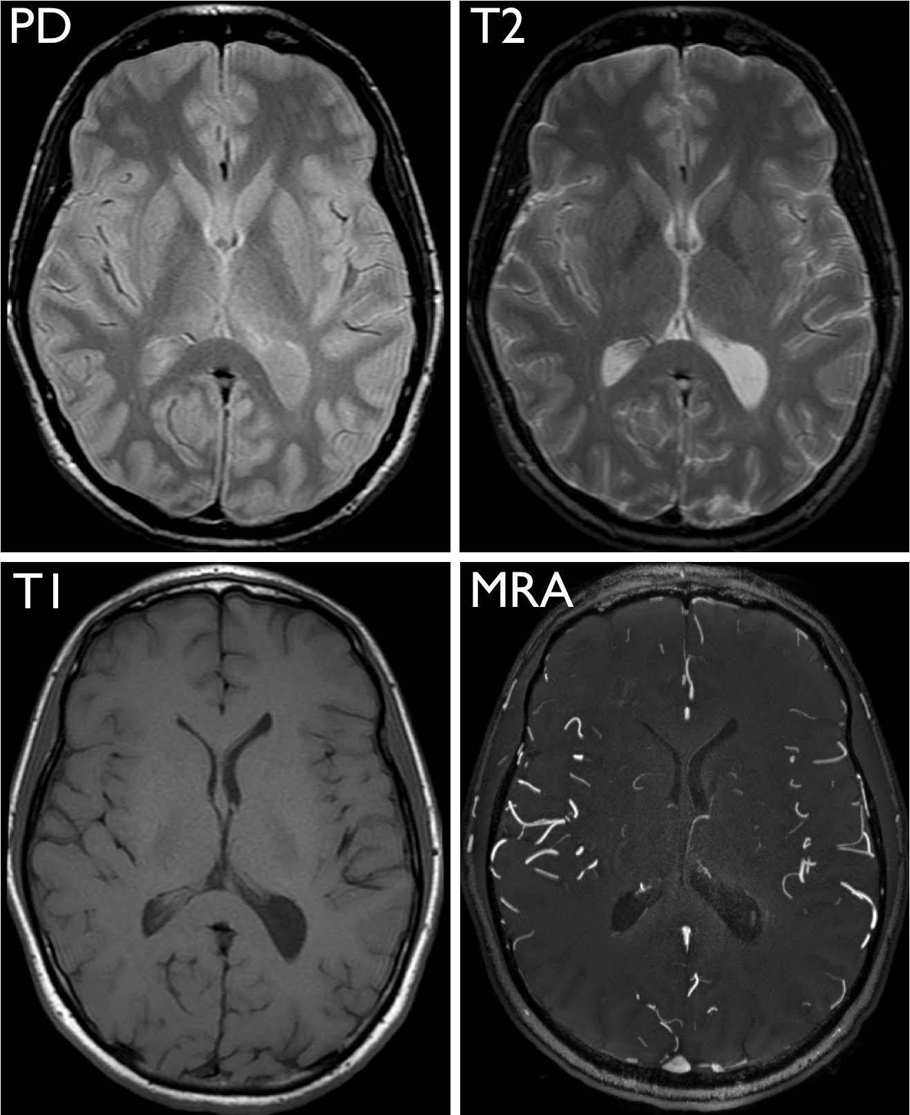
Exploring the Brain How Are Brain Images Made with MRI? UCSF Radiology

MRI anatomy brain axial image 6 Mri brain, Brain anatomy, Mri

MRI scan Morton & Partners Radiologists
Investigation of these patients includes thorough clinical examination, audiological evaluation and frequently Magnetic Resonance Imaging (MRI) of the Internal Auditory Meatus (IAM), cerebellopontine angle (CPA) and brain. MRI scanning is a well-established, cost-effective investigation for these patients [1-3]. Only a small percentage of.. MRI is also a tomographic imaging modality, in that it produces two-dimensional images that consist of individual slices of the brain. Images in MRI need not be acquired transaxially, and the table or scanner does not move to cover different slices in the brain. Rather, images can be obtained in any plane through the head by electronically.